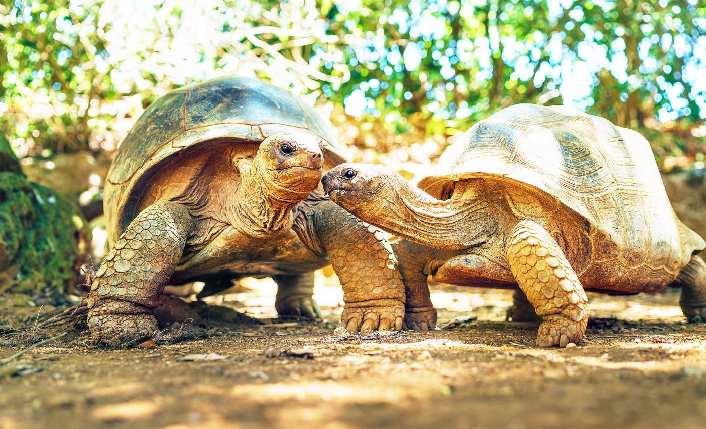
Turtles have genomes unlike any other animal studied so far
Turtles are fascinating reptiles known for their distinctive shells, which serve as both armor and protection. They’ve been around for over 200 million years, making them some of the oldest living creatures on Earth.
You can find turtles in a variety of environments, from the depths of the ocean to freshwater ponds and even on land. Their ability to adapt to different habitats has allowed them to thrive across the globe, with hundreds of species showcasing a wide range of sizes, colors, and behaviors.
And where does this impressive ability to adapt come from? Apparently, it’s their genome, which is proving to be very different from that of most other animals studied so far.
Insights into genetic blueprints
DNA molecules, with their long strands of nucleotides, carry extensive genetic data essential for life. How this genetic blueprint is stored in cells influences how it is read and used.
During cell division, DNA is tightly coiled around proteins, forming chromatin structures within chromosomes.
After division, the chromosomes relax, and chromatin loosens, creating unique folding and looping patterns that determine which genes are activated or repressed.
New research led by Iowa State University offers insights into these processes, with implications for understanding evolution and potentially aiding biomedical research.
Study senior author Nicole Valenzuela is a professor of ecology, evolution, and organismal biology at Iowa State University.
“The three-dimensional structure of the chromatin when it’s folded matters for gene regulation. Where chromatin physically sits in the nucleus matters,” said Valenzuela.
“Chromosome folding remains a bit of a black box. We’ve learned a lot about it, but it’s still just the tip of the iceberg.”
Role of chromatin in gene regulation
In the post-division interphase stage of the cell cycle, the chromatin’s shape and location impact gene expression by bringing distant DNA regions into close contact.
For example, enhancers (DNA segments that promote gene expression) and gene promoters (regions that initiate transcription) can interact more readily in open, accessible regions of chromatin.
On the other hand, DNA in tightly packed chromatin regions remains less active.
Through analyzing these contact points, researchers have developed models to map chromatin configurations across various species, such as humans, mice, birds, and more recently, turtles.
Unique discovery in turtle genomes
In a recent paper published in the journal Genome Research, Valenzuela’s team described the chromatin arrangement in the genomes of two turtle species, the spiny softshell and northern giant musk turtles, uncovering a structure previously unobserved in other organisms.
Chromosomes have two essential elements: the centromere, a constriction point, and telomeres, which cap each chromosome end. In humans, chromosomes occupy separate regions in the nucleus.
However, in some animals, like marsupials, centromeres cluster, while in birds, telomeres come together.
Turtles, interestingly, are the first species where both telomeres and centromeres align near each other, suggesting a unique chromatin arrangement.
“It’s possible this is the ancestral condition of amniotes, from which mammals, birds, and reptiles evolved in different patterns,” Valenzuela noted.
“The turtles may be showing us what existed at the beginning, shedding light on the evolution of vertebrate genomes.”
Turtle genomes and biomedical science
Uncovering the chromatin structure in turtles and how it adapts to environmental changes could be valuable for biomedical science.
For example, some turtle species can survive weeks without oxygen, which could have implications for treating strokes.
Similarly, understanding how certain turtles withstand extreme cold might inform techniques for cryogenic preservation of human tissues.
“We want to understand more about why different lineages are different in some aspects and why they’re the same in others, what parts we share and what parts differ,” Valenzuela explained.
“If we can reconstruct the evolutionary history of the changes that have taken place, we will be able to tell much more about how the differences in the packaging of the DNA and the folding of chromosomes might be affecting the traits we’re interested in, how genes are regulated, and how vertebrate genomes evolve.”
Additionally, understanding turtles’ chromatin structure could aid conservation efforts, offering insights into how environmental changes impact their biology and potential adaptations.
Future research directions
Valenzuela’s lab plans to extend their research to study more turtle species, adding to the current work on spiny softshell and northern giant musk turtles.
Her team has already gathered data on four additional species, aiming to see if similar chromatin structures appear in genomes across different turtle lineages.
The researchers are also interested in comparing turtle chromatin organization to that of other reptiles, such as crocodiles, lizards, and snakes, to explore whether they share structural patterns.
To gain deeper insights, the team will study liver organoids – lab-grown tissue mimics they have developed for three turtle species.
These organoids offer a simplified view of liver tissue, allowing the researchers to observe how chromatin folding might influence cellular function.
Future work will include advanced mapping techniques that provide high-resolution chromatin data, helping to clarify how 3-D chromatin structures change over time and under various environmental conditions.
“To really do genotype-to-phenotype mapping, we have to get to this level of complexity,” Valenzuela concluded.
—–
Like what you read? Subscribe to our newsletter for engaging articles, exclusive content, and the latest updates.
Check us out on EarthSnap, a free app brought to you by Eric Ralls and Earth.com.
—–


