How do plants respond to specific stress situations?
When it comes to survival, plants face a significant challenge compared to many other living organisms: they cannot move to escape predators, pathogens, heat stress, or other unfavorable environmental conditions.
To cope with these threats, plants have evolved various strategies triggered by specific environmental signals. It has long been known that changes in intracellular calcium concentration play a key role in processing these signals.
However, alongside these calcium changes, shifts in the cell’s membrane potential have also been suspected to act as signal transmitters.
Now, researchers from the Departments of Neurophysiology, Pharmaceutical Biology, and Botany at Julius-Maximilians-Universität Würzburg (JMU) have explored the relationship between calcium and membrane potential in greater depth.
Plants’ response to stress
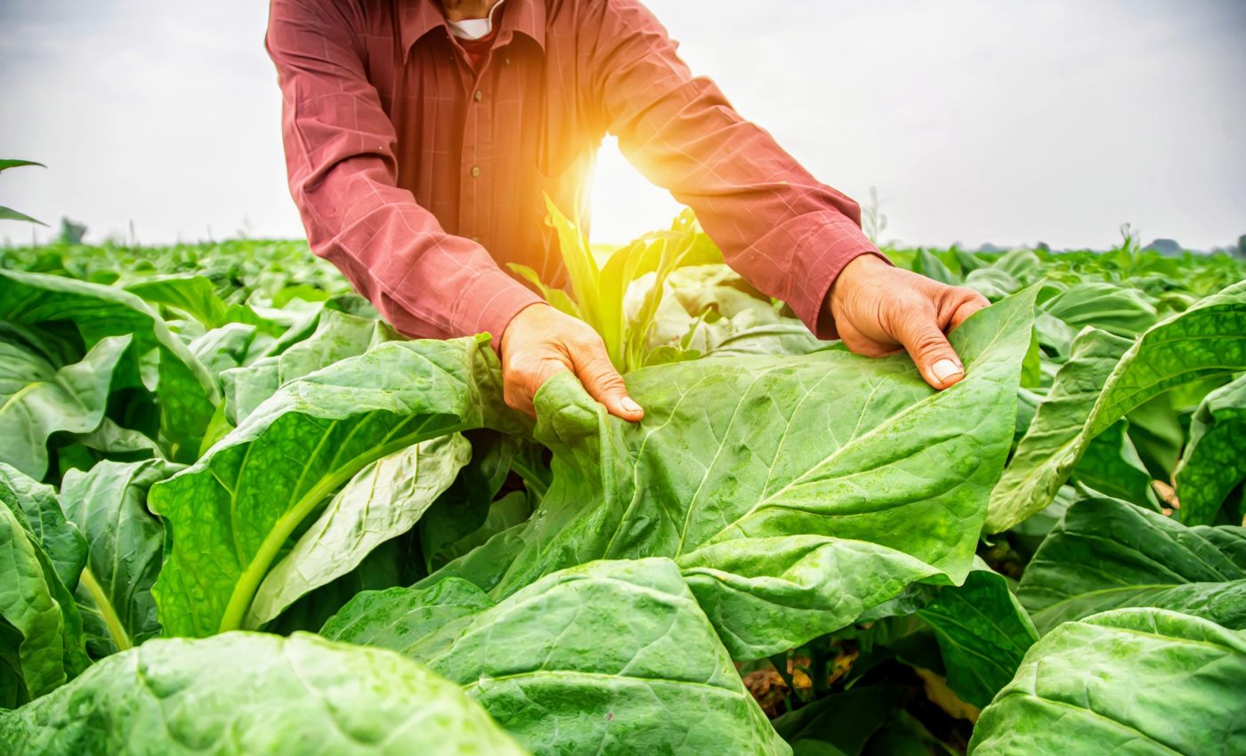
In their study, the research teams worked with tobacco plants that were engineered to carry ion channels that could be specifically activated by light.
Over 20 years ago, scientists Peter Hegemann, Georg Nagel, and Ernst Bamberg pioneered the field of optogenetics with their discovery and characterization of light-activated ion channels, known as channelrhodopsins.
Using these light-sensitive proteins, which are derived from algae and microorganisms, the JMU researchers were able to experimentally determine whether the influx of calcium ions or the depolarization of the cell membrane, mediated by anion efflux, was crucial for the plant’s response to specific stress situations.
However, considerable preparatory work was required before they could conduct these experiments.
Optimizing the use of channelrhodopsins
Channelrhodopsins, ion channels equipped with a rhodopsin-based light switch, revolutionized neuroscience by enabling the light-controlled study of neuronal networks.
The application of channelrhodopsins in plant research became feasible only 20 years later, thanks to close collaboration between Georg Nagel’s group at the Institute of Physiology at JMU and plant researchers from the Würzburg Chairs of Botany 1, 2, and Pharmaceutical Biology.
In 2021, Nagel’s group, along with Dr. Kai Konrad, a group leader at the JMU Chair of Professor Rainer Hedrich Botany 1, published a method to optimize the use of channelrhodopsins in plants by overcoming three major challenges.
Successful expression of channelrhodopsins
Study co-author Shiqiang Gao, a “rhodopsin engineer” from the Optogenetics lab in the Department of Neurophysiology at JMU, explained the first challenge.
“Like all rhodopsins, including those in our eyes, channelrhodopsins require the small molecule retinal, also known as vitamin A, to absorb light. We humans get retinal mainly from beta-carotene, the provitamin A. However, land plants do not contain retinal, but they do have plenty of beta-carotene,” said Gao.
In 2021, Gao succeeded in combining the expression of channelrhodopsins with the production of retinal from beta-carotene in plant cells. This breakthrough allowed the development of tobacco plants with a high retinal content and successful expression of channelrhodopsins.
Dr. Markus Krischke from the Metabolomics Core Unit in the Department of Pharmaceutical Biology, headed by Professor Martin Müller at JMU Würzburg, confirmed the high retinal content of various transgenic tobacco plants.
Comparable transgenic tobacco plants were produced for the recently published study by Dr. Meiqi Ding from the Department of Botany 1, under the direction of plant physiologist and plant signal processing expert Dr. Kai Konrad, from Professor Rainer Hedrich’s group at Botany 1.
The challenge of light activation
The second challenge involved light activation. “Most rhodopsins are activated by blue or green light. However, these colors are components of white light,” explained Nagel.
This meant the tobacco plants could not be grown in a greenhouse or under standard artificial white light conditions, as this would inadvertently activate the rhodopsins.
Instead, the plants had to be grown in special growth chambers with red LED light, which is photosynthetically useful and does not trigger rhodopsin activation.
Tests under various growth conditions revealed that tobacco develops healthily and unchanged under red light compared to greenhouse conditions, noted Konrad.
Channelrhodopsins in tobacco cells
The third challenge was the difficulty in expressing channelrhodopsins in tobacco cells. In 2021, the Würzburg research team successfully expressed the light-activated anion channel GtACR1 in tobacco plant cells.
As a result, Nagel’s team was able to develop various channelrhodopsins optimized for calcium ion permeability.
Ultimately, Shiqiang Gao and Shang Yang, both members of Nagel’s group, succeeded in developing an advanced calcium-conducting channelrhodopsin, named XXM 2.0, specifically for targeted expression in tobacco plants.
This marked a significant breakthrough: “The successful expression of channelrhodopsins with different ion selectivities in plant cells allows us to compare different ion signals alongside the electrical signal, known as depolarization,” Meiqi Ding explained.
She used the calcium-conducting channelrhodopsin XXM 2.0 and the light-activated anion channel GtACR1 to investigate the various ion signaling pathways in tobacco.
Optogenetic tobacco plants
These newly created “optogenetic” tobacco plants enabled the researchers to answer the question of whether calcium influx or membrane depolarization is more critical for the plant’s response to a specific stress situation. “The answer was clear,” said Konrad.
“After activation of the anion channel, the leaves wilted and showed the typical plant response to drought; the plant hormone abscisic acid (ABA) was produced, and gene expression was increased to protect against dehydration,” Ding added.
“However, in the plants with the calcium channel, there was no change in ABA levels after optogenetic stimulation.”
“Instead, the plants produced signal molecules and plant hormones to trigger defense mechanisms against predators, as evidenced by white spots on the leaves,” Konrad said.
A new era in plant research
Dr. Sönke Scherzer from Professor Hedrich’s chair demonstrated through direct measurements that reactive oxygen species (ROS) were released during this process.
Dirk Becker and Rainer Hedrich at Botany 1 designed an experimental approach using transcriptomic and bioinformatic analysis to support the working hypothesis.
The researchers are convinced that this study marks the beginning of a new era in plant research. They believe that with the help of various rhodopsins, plant signaling pathways can now be more thoroughly “illuminated.”
The study is published in the journal Nature.
—–
Like what you read? Subscribe to our newsletter for engaging articles, exclusive content, and the latest updates.
Check us out on EarthSnap, a free app brought to you by Eric Ralls and Earth.com.
—–


